LLPS (Liquid-liquid phase separation)
Liquid-liquid phase separation (LLPS) has become a focal point in recent biological studies, describing a phenomenon where biomolecules surpass solubility, resulting in the condensation and separation of these molecules into liquid-like droplets. In the case of a solution containing macromolecules such as proteins or nucleic acids, undergoing LLPS leads to condensation into a dense liquid droplet phase alongside a dilute phase. Holotomography proves valuable for observing this phase transition without requiring additional labeling. The refractive index change during the phase transition allows for real-time observation of the process. Moreover, quantitative analysis is feasible using refractive indices, enabling measurements of volume, mass, protein concentration, and sphericity.
Features
Discover LLPS with HT
-
Phase-separated liquid droplet study
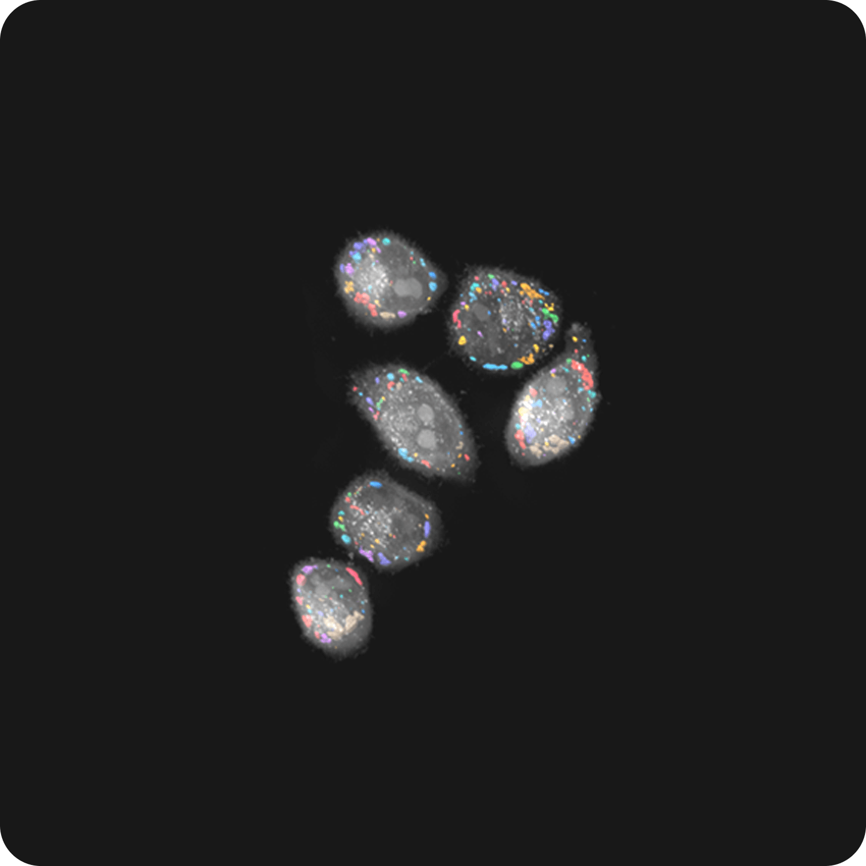
In a 2023 study featured in Nature Cell Biology, HT technology emerged as one of the key players, confirming the influence of OASL phase condensation in nucleating RIPK3 and promoting amyloid fibril formation (Lee et al. 2023). RI enables the characterization of membraneless OASL droplets in a label-free and noninvasive manner. Meanwhile, the combination of 3D HT and fluorescence imaging provided quantitative measurements of the morphological and biochemical properties of the droplets. It also revealed the increasing nucleation of RIPK3 over time as OASL droplets grew in size and concentration.
-
Phase separation in the nucleus
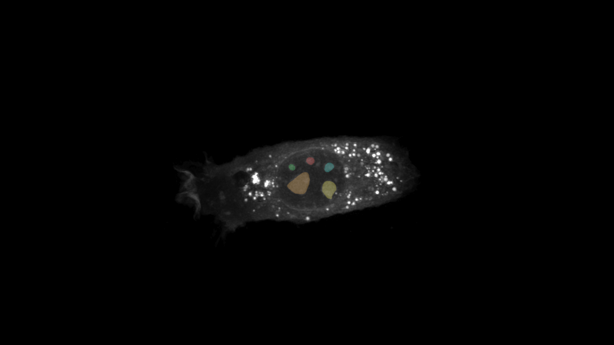
In a 2023 study published in Nature Communications, HT imaging imparts crucial insights into biomolecular condensates and intracellular phase behavior (Kim et al., 2023). HT technology excels in distinguishing condensates based on mass concentration, categorizing them into distinct groups. Notably, high-density condensates, including nucleosomes and heterochromatin, sharply contrast with low-density counterparts like nuclear speckles and stress granules. The integration of fluorescence further enhances HT's analytical capabilities. When combined, HT and fluorescence collectively reveal influential factors impacting low-density condensates, including the roles of RNA and organelles. This holistic approach allowed scientists to comprehensively grasp the intricate dynamics within the cellular microenvironment.
Selected publications
-
HT technology emerged as one of the key players, confirming the influence of OASL phase condensation in nucleating RIPK3 and promoting amyloid fibril formation. RI enables the characterization of membraneless OASL droplets in a label-free and noninvasive manner. Meanwhile, the combination of 3D HT and fluorescence imaging provided quantitative measurements of the morphological and biochemical properties of the droplets and revealed the increasing nucleation of RIPK3 over time.
-
HT technology excels in distinguishing condensates based on mass concentration, categorizing them into distinct groups of high-density condensates, such as nucleosomes and heterochromatin, and low-density condensates, such as nuclear speckles and stress granules. The integration of fluorescence further enhances HT's analytical capabilities. When combined, HT and fluorescence collectively reveal influential factors impacting low-density condensates, including the roles of RNA and organelles.