Cell biology
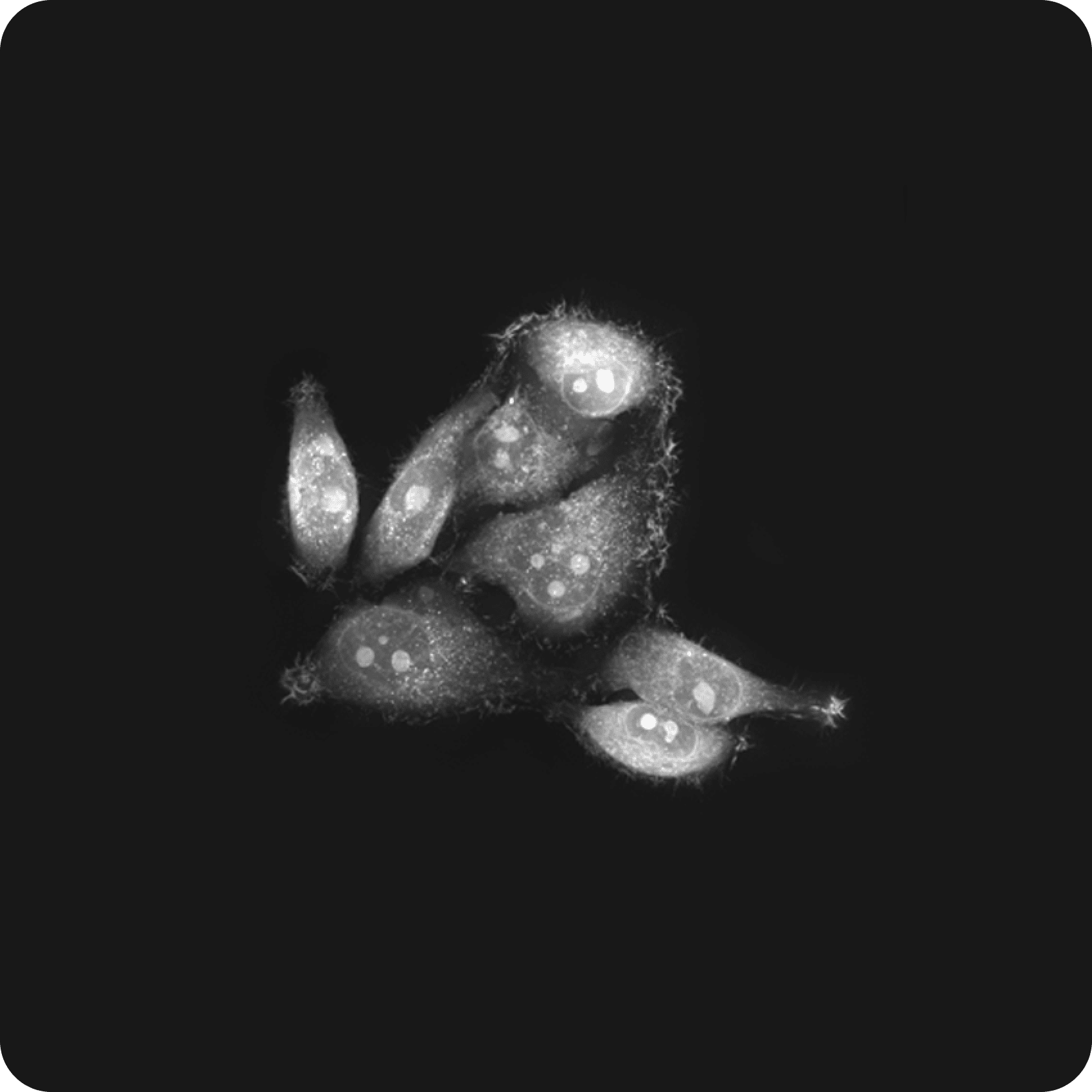
Cell biology, a crucial domain in biological research, aims to unravel the intricacies of cell structures, their functions, and the diverse interactions occurring between cells. Holotomography (HT) emerges as a powerful tool in this field by not only visualizing the transparent cells at their native state label-free but also revealing subcellular structures like nuclei, nucleoli, lipid droplets, and mitochondria in high resolution. The label-free approach significantly advances our understanding of cellular dynamics.
Holotomography provides high-content 3D visualization and furnishes quantitative data for individual cells and their intracellular structures. 3D cell images make it possible to understand differences in cell characteristics depending on cell volume, surface area, and three-dimensional shape that cannot be seen in 2D images. Because of the high resolution, the inside of the cell can be observed clearly and in detail. The growth and development of lipid droplets can be observed in the adipogenesis process. In addition, pinocytosis, where vacuoles enter the cell membrane, and mitochondrial elongation and fragmentation can be observed in real time without staining. Interested regions can be segmented by refractive index (RI) combined with morphological characteristics and measured using the known Refractive Index Increment (RII). Through the quantitative analysis, HT images offer physical quantities such as mean RI, dry mass, and concentration, alongside shape measurements like area, length, volume, surface area, projected area, circularity, sphericity, and center of mass. Leveraging the mean RI, which represents material characteristics, along with dry mass and concentration, facilitates an in-depth analysis.
The morphological and biophysical information of each segmented cell's internal structure provides the exploration of various life phenomena, including cell interaction, inter-communication, cell migration, metastasis, and apoptosis.This comprehensive approach enables a thorough analysis of material properties and cellular dynamics based on HT data. Holotomography stands out as a potent tool in advancing our understanding of cell biology, making significant contributions to the ongoing developments in this scientific domain.
Features
Discover cell biology with HT
-
Live cell imaging
Live cell imaging offers real-time insights into cellular responses to diverse stimuli by capturing and recording complex biological processes within living cells. Holotomography enhances this capability by enabling immediate examination without the need for pre-treatment processes and facilitating continuous monitoring of cellular phenomena overtime. This methodology proves invaluable in studying fundamental cellular processes, including cell division, migration, cell interaction, signaling pathways, and cellular responses to drug-induced or environmentally induced changes. The broad applications of this advanced imaging technique span various scientific domains, prominently featuring in cell biology, pharmacology, and diverse biomedical research areas. Its widespread use significantly contributes to a comprehensive understanding of cell function within the natural and dynamic living environment.
-
Lipid droplet biogenesis
Lipid droplets, crucial energy storage organelles for cells, play vital roles in various physiological processes, including those observed in hepatocytes and immune cells. Conventional imaging methods often rely on special dyes like Nile red or BODIFY to visualize transparent lipid droplets, a technique typically limited to fixed cells. Furthermore, dyes for living cells possess relatively low molecular specificity, introducing challenges such as light bleaching and phototoxicity during extended imaging sessions.
HT presents a distinctive advantage by measuring the average refractive index (RI) of each pixel. With the RI of lipid droplets being 1.38 or higher, HT enables effective compartmentalization of lipid droplets in the imaging process.
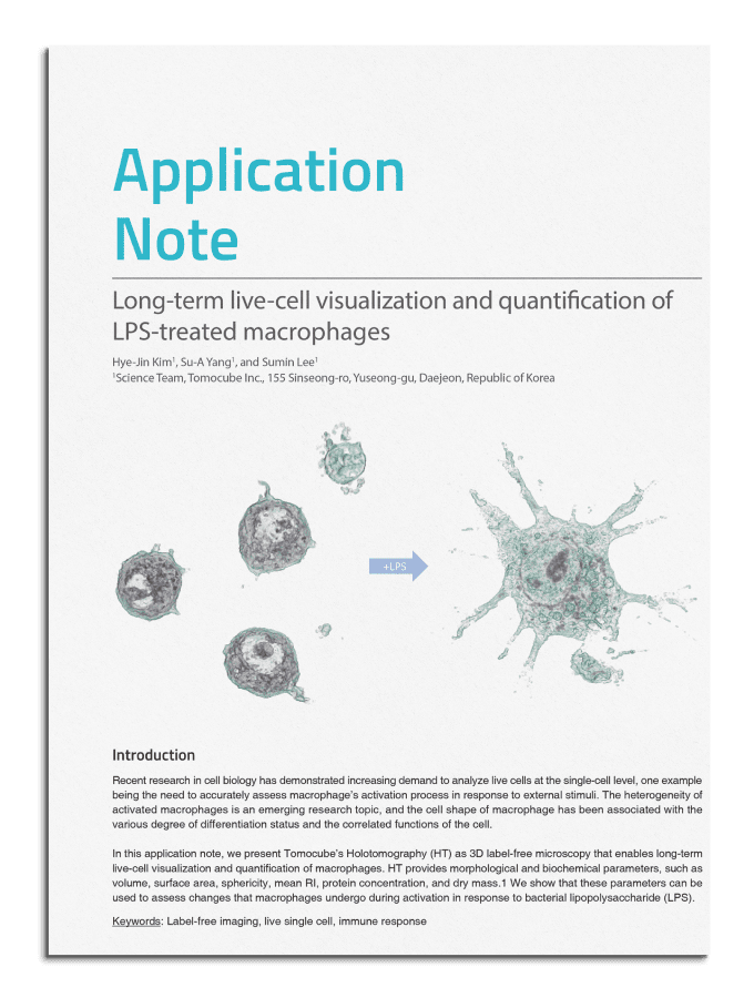
In a noteworthy study published in ACS Nano in 2020, researchers quantitatively analyzed the suppression of lipid droplet production over time in macrophages and foam cells—key players in arteriosclerosis—when subjected to drug treatments. The analysis involved quantifying various parameters, including the number, volume, and dry mass of lipid droplets, with a comparative assessment between different treatments, particularly in response to nanomaterial treatment (Park et al., 2020). Another study highlighted the potential of HT-X1 products by extensively observing a 42-day process wherein human knee chondrocytes underwent reverse differentiation to form dedifferentiated adipocytes (DFAT) and subsequently re-differentiated into mature adipocytes (Jeong et al., 2024). This application of HT technology allows for detailed and dynamic investigations into lipid droplet dynamics and related cellular processes.
-
Cell-in-cell structures
Cell-in-cell (CIC) structures, where one cell engulfs another, creating a unique cellular arrangement, offer insights into cellular behaviors and interactions. HT excels in rapidly capturing three-dimensional shapes without staining, making it ideal for observing diverse cell interactions in their native state. In a recent study, the CIC phenomenon, where NK cells entered cancer cells, was identified using HT imaging (Choe et al., 2022). Utilizing label-free HT observation of CIC structures between cancer and NK cells, the study focused on heterotypic cell segmentation, facilitating a further analysis of its contribution to drug resistance. Continuous monitoring of the time-lapse process of cancer cell death added temporal insights. Incorporating correlative fluorescence imaging aided in distinguishing different cell types, enhancing the understanding of dynamics within cell-in-cell structures.
-
Cell migration
Cell migration assays in a controlled environment often employ live cell timelapse imaging to study dynamic cellular movements, providing insights into the mechanisms underlying physiological activities like wound healing or cancer cell metastasis. HT emerges as a crucial tool for monitoring fundamental cell behaviors, encompassing proliferation, migration, and spreading.
Migrasomes, formed in migrating cells, are gaining renewed attention in the realm of extracellular vesicles. In a recent study, HT was employed to observe that TNF alpha increased the formation of migrasomes (Gagat et al. 2021). HT played a crucial role in correlating the 3D morphology with localization in correlative fluorescence imaging, enabling stereoscopic observation of migrasomes.
Resources


Selected publications
-
Researchers utilized HT for label-free, 3D, timelapse monitoring of living foam cells to assess the therapeutic impact of designed nanodrugs on lipid droplet production in macrophages and foam cells - a crucial feature in arteriosclerosis. Machine-learning-based image analysis was also employed for single-cell level therapeutic evaluation.
-
HT was used to assess the uptake of hyaluronic acid-based nanoparticles (FHA-NPs) and its effect on cell morphology and organelle dynamics in lung carcinoma epithelial cells. 3D refractive index images helped confirm the important role of lipid metabolism in ferroptosis through the evident accumulation of lipid droplets/cytoplasmic lysosomes in treated cells.