Organoids
See Your Organoids Live, Then Take the Same Sample Into Your Next Assay.
Cells behave differently in three dimensions. The neighbors they touch, the matrix they sit in, and the gradients that build across a 3D structure all shape how a tissue forms, how disease takes hold, and how it responds to a drug. That context is what a single cell or a flat monolayer cannot give you, and it is why disease modeling and compound screening increasingly run on organoids instead.
But the very thing that makes an organoid worth growing, its living 3D structure, is also what makes it hard to see. Image it the 2D way and you collect z-slice after z-slice, then struggle to turn those stacks into something you can actually analyze. The deeper problem is not fluorescence itself, it is having no way to see structure without it. Stains have to penetrate an organoid embedded in 3D gel, so your data is only as good as your staining efficiency. Dense organoids make that harder still, and optimizing a staining protocol can eat up real time before the real question even gets asked.
Holotomography removes those bottlenecks. To see and measure an organoid's 3D structure, you no longer stain it or engineer in a fluorescent reporter. HT reads refractive index and reconstructs the whole intact, living organoid as a single 3D volume, quantifying shape, volume, dry mass, and density directly. There is no penetration to fight, no staining protocol to optimize, no stack to reassemble, and no label-induced artifacts to second-guess.
Fluorescence becomes a choice, not a requirement. When a question truly needs molecular identity, correlative fluorescence overlays it on the same living organoid, registered against the full label-free 3D structure. And because label-free imaging is non-destructive and low in phototoxicity, the same organoid can stay in culture for sequencing, staining, or functional assays.
Features
-

- Label-free live cell imaging
- Holotomography (HT) imaging eliminates the need for labeling or dyeing, therefore minimizing artificial manipulation effects and simplifying sample preparation. This approach allows the observation of samples in their natural state, providing more reliable and relevant information.
-
- High-resolution 3D imaging
- HT provides high-content, high-resolution, three-dimensional images, allowing researchers to capture detailed information about cellular structures. This high level of detail is crucial for studying cellular morphology and dynamics.
-
- Long-term timelapse imaging
- HT facilitates continuous monitoring of cellular phenomena over extended time frames. This feature is beneficial for studying processes that unfold over hours, days, or even weeks, providing a comprehensive understanding of long-term cellular behavior.
-

- Quantitative analysis
- HT allows for quantitative analysis of cellular parameters. It can measure physical quantities such as mean refractive index,dry mass, and concentration, as well as various shape measurements like area,length, volume, and surface area.
Discover Organoids with HT
-
Organoids: Miniature Organs for Advanced Research
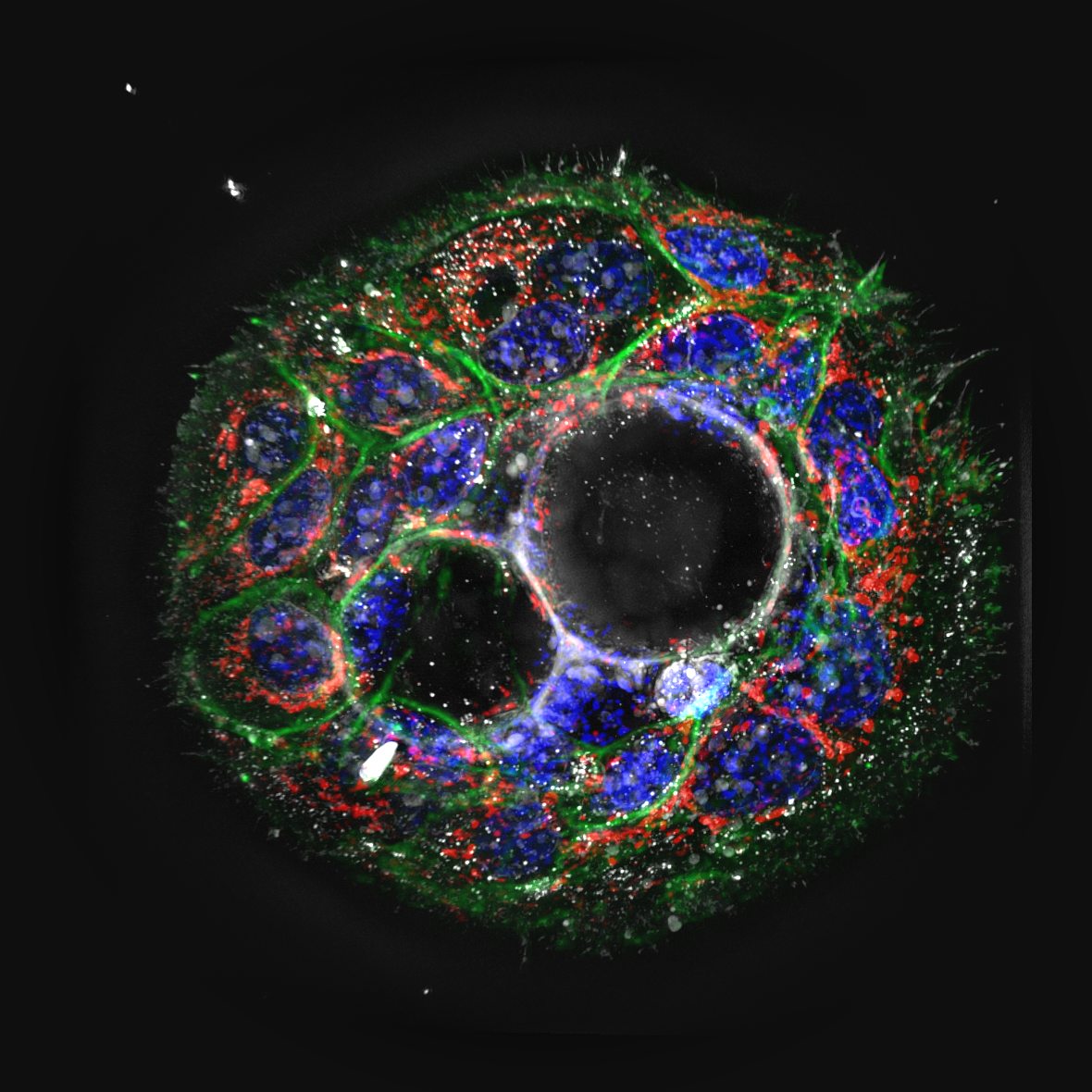
Research on organoids, the 3D cell aggregates replicating organ structures and functions, benefits significantly from the detailed and noninvasive imaging capabilities of Holotomography (HT). HT efficiently captures the dynamic behavior of cells within living organoids, including mitotic events, cyst formation, and crypt budding. Real-time, high-resolution observations with available quantitative measurements facilitated by HT are crucial for exploring the complex dynamics and morphology of the organoids.
In combination with the HT-ready 96-well plate, which supports a broad spectrum of cell types from 2D to 3D cultures, the HT-X1 is an ideal tool for leveraging the potential of organoids in drug development. It enables high-throughput screenings for rapid assessment of drug response and its toxicity on patient-derived tissues.
A recent study highlighted a successful example of such efforts, demonstrating how HT has set a new standard for comprehensive and rigorous statistical assessments for the biological studies of organoids (Lee et al., 2023). With HT, intricate subcellular structures of individual cells in small intestinal organoids were observed in intricate detail. Long-term monitoring of various biological processes within the specimen, including cell mitosis, migration, apoptosis, and other subcellular dynamics, was achieved effortlessly without the need for labeling or other time-consuming preparations.
Additionally, the observation of morphological changes was combined with quantitative parameters, including organoid volume, protein concentration, and mass, to effectively and comprehensively evaluate the response of the organoids to chemotherapy drug treatments. The study suggested that the Tomocube HT-X1 imaging system is an indispensable tool for organoid-based pharmacological screening applications. -
Microphysiology Systems & Organ-on-a-Chip
Bridging the Gap Between in vitro and in vivo
Microphysiology systems (MPS) and organ-on-a-chip devices represent the next evolution in physiologically relevant cell culture models. These microfluidic platforms recreate tissue-tissue interfaces, mechanical forces, and fluid flow conditions that are impossible to achieve in static cultures.
Why do traditional imaging methods fall short for MPS?
Organ-on-a-chip devices present unique imaging requirements:- Continuous flow systems cannot tolerate fixation or staining protocols
- Scaffold materials often exhibit autofluorescence
- Multi-layer architectures require true 3D imaging capability
- Real-time monitoring is essential for dynamic studies
What advantages does holotomography offer?
Holotomography is uniquely suited for MPS imaging because it requires no sample preparation that would disrupt the carefully controlled microenvironment:- Non-invasive observation: Image cells on collagen scaffolds and hydrogel matrices without interference
- Real-time monitoring: Track cellular responses to flow conditions and chemical gradients
- Quantitative analysis: Measure cell morphology, proliferation, and viability continuously
- Multi-day experiments: Monitor device function over extended culture periods
Common applications include:
- Liver-on-a-chip for hepatotoxicity screening
- Gut-on-a-chip for barrier function studies
- Lung-on-a-chip for inhalation toxicology
- Vascular chips for endothelial research
The combination of label-free imaging and quantitative 3D analysis makes holotomography an ideal tool for validating and characterizing microphysiology systems during development and application.
Resources
-
Live Organoid Dynamics | HT-X1, TomoAnalysis, Paneth Cell
Download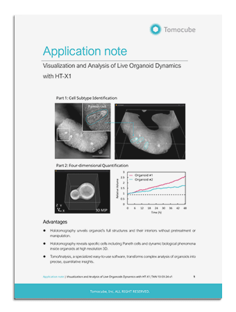
-
Dynamic Live Cell Imaging in Microfluidic Chips
Download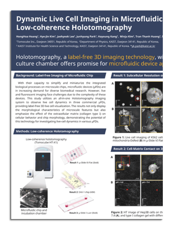
-
Investigating Cellular Dynamics of Live Organoids
Download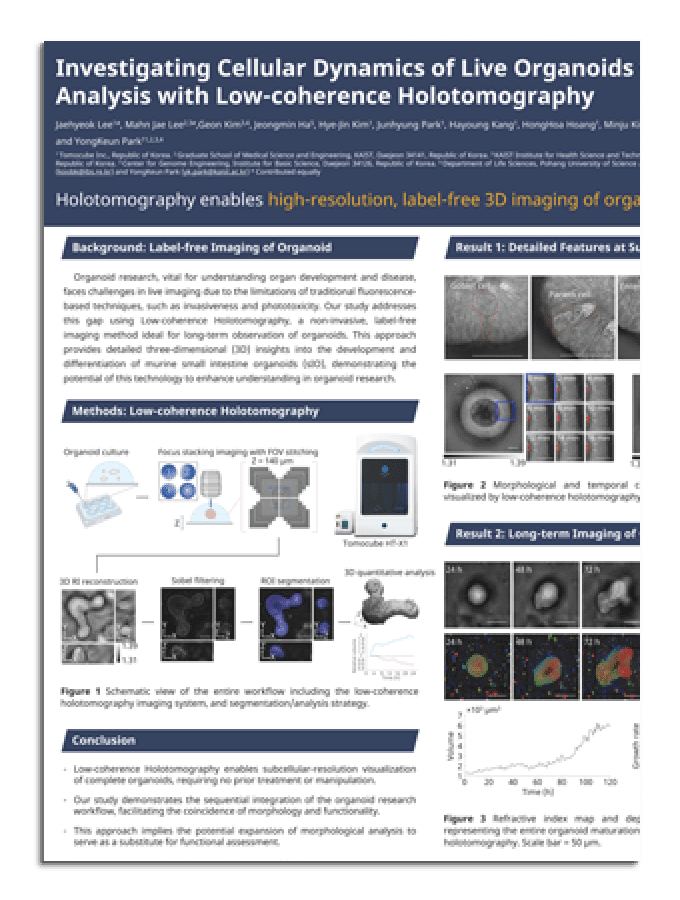
-
Advanced Visualization Strategies of Organoids
Download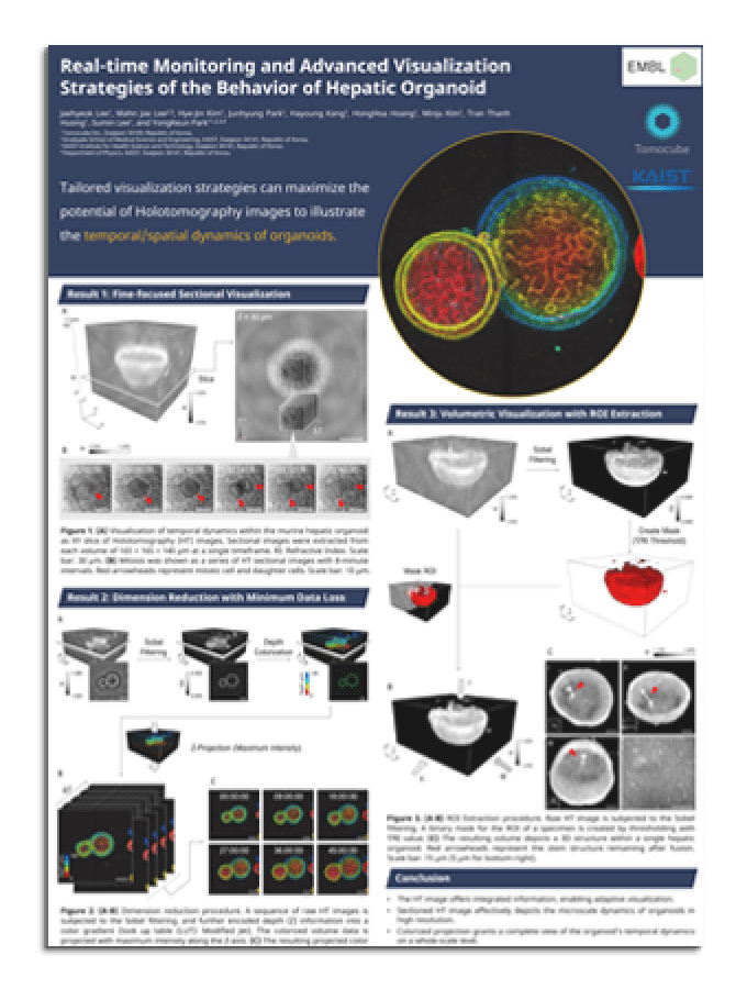
-
3D Imaging of Neurons and Brain Structures
Download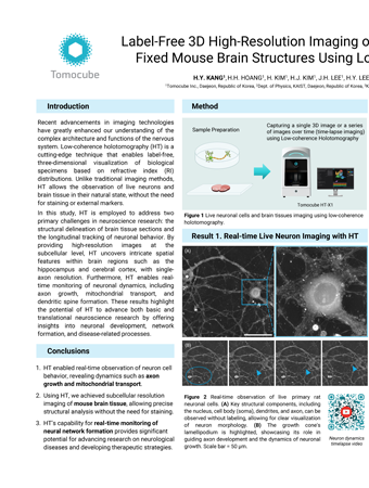
-
AI-Assisted Lipid Droplet Dynamics Analysis in Live Organoids
Download
-
AI-Enhanced Segmentation of 3D Cell Structures
Download