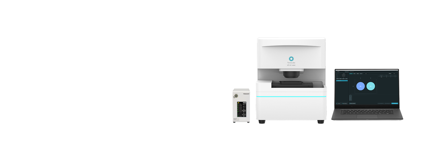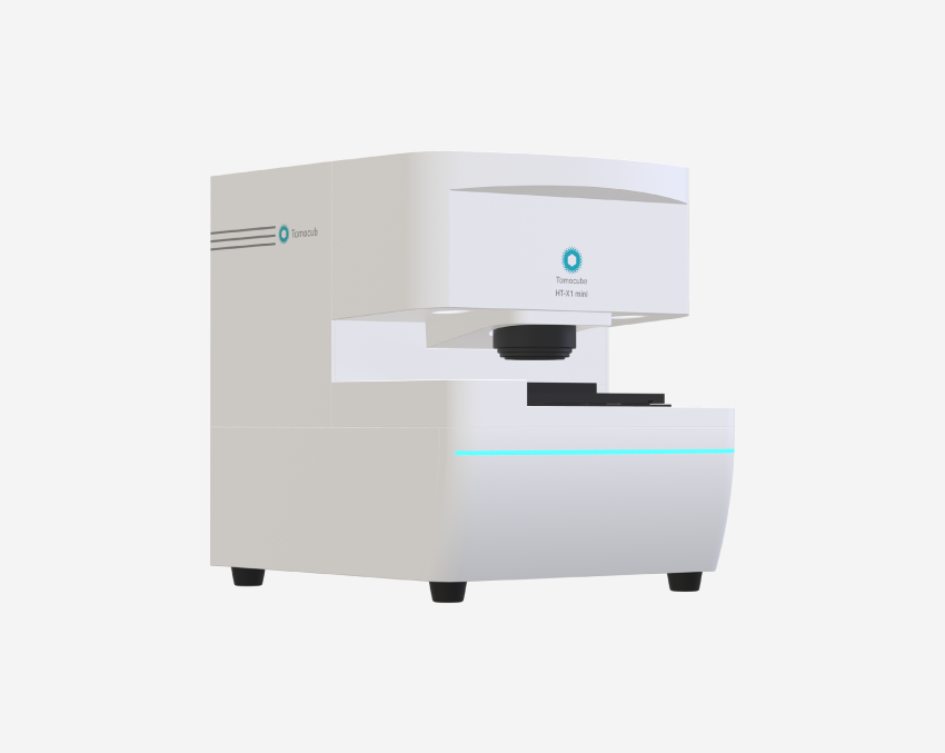
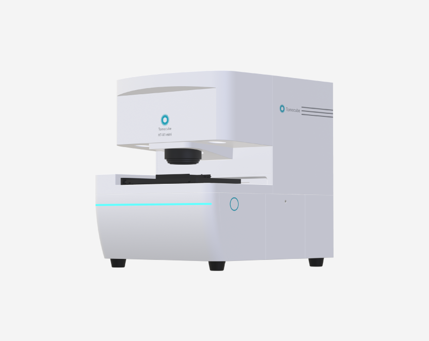
HT-X1™ mini
A compact design with flexible configuration, delivering uncompromised quality and making advanced Holotomography imaging accessible for every lab.
The HT-X1™ mini makes advanced holotomography accessible to all. Compact, intuitive, and ready for any lab bench, it transforms everyday research into breakthrough 3D imaging. Its portable, space-saving design allows effortless installation even in limited environments such as cell culture rooms, clinical labs, or field labs.
Powered by the same 2nd generation holotomography technology as the HT-X1 series, it delivers high-resolution, label-free 3D imaging of live cells. Fully compatible with TomoAnalysis™, the HT-X1™ mini ensures the same high-quality image analysis as the HT-X1 series, providing seamless continuity in research.
The HT-X1™ mini is built to grow with your research. Start with the essentials, then expand capabilities with four upgrade modules — fluorescence imaging, environmental incubation, laser-based autofocus, and multi-wavelength illumination. This modular flexibility lowers initial investment barriers while enabling your system to adapt to new experimental needs — supporting everything from routine cell culture checks to complex, high-content 3D imaging.
Features
-
- Compact design
- Small footprint fits seamlessly into most lab environments without disrupting workflow.
-
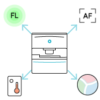
- Expandable functionality
- Upgradeable with fluorescence, incubator, laser-based autofocus, and multi-wavelength illumination for flexible research needs.
-
- Uncompromised Quality
- Delivers the same consistent, reliable holotomography image as the HT-X1 Series.
-

- Analysis compatibility
- Fully compatible with TomoAnalysis™ software, including AI-powered quantitative analysis tools.
User benefits
-
Compact Design with Expandability
The HT-X1™ mini is an accessible system that can be easily introduced into any individual laboratory, from small research groups to multidiscipline institutions. It combines a compact footprint with flexible expandability, allowing researchers to start simple and add advanced modules — such as fluorescence imaging, long-term time-lapse with environmental control, laser-based autofocus, and additional wavelengths — as their research evolves. This makes it a future-proof model that grows with the needs of the lab while maintaining ease of use and space efficiency. With this adaptability, researchers can perform label-free 3D live-cell imaging in virtually any setting, including cell culture rooms and shared facilities.
-
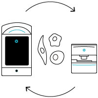
Everyday Access to Advanced Imaging
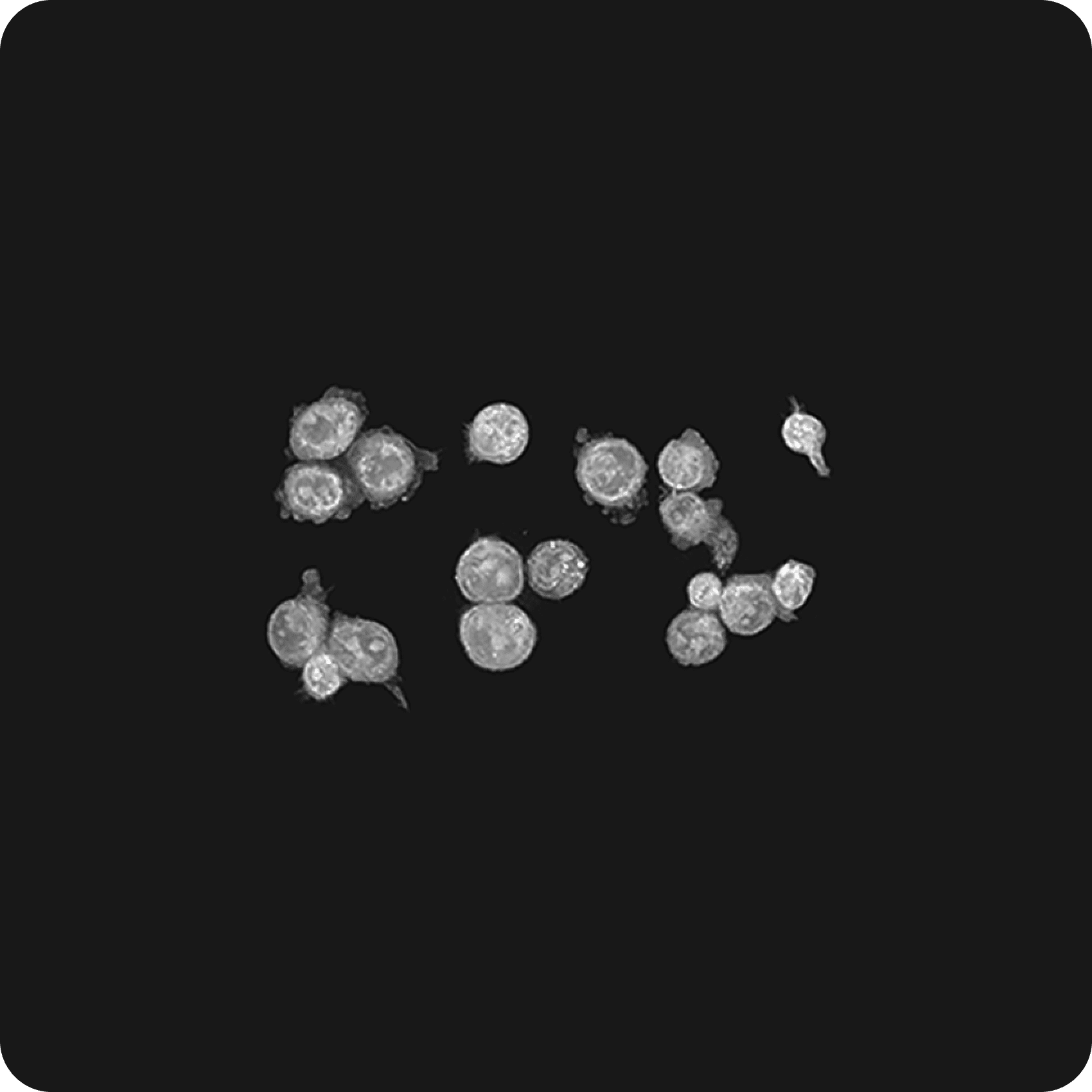
The HT-X1™ mini integrates advanced holotomography into everyday research workflows with its compact footprint and intuitive operation. Designed for space-limited environments — from cell culture rooms to mobile field laboratories — it can be positioned in routine experimental areas for convenient access to imaging within your workflow. This proximity enables quick checks such as cell confluency monitoring. With holotomography’s label-free capability, researchers can observe cells in their natural state, track cell count changes over time, and continue culturing the same samples — integrating everyday tasks into the imaging workflow while maintaining sample integrity.

-
Fully Compatible with TomoAnalysis™
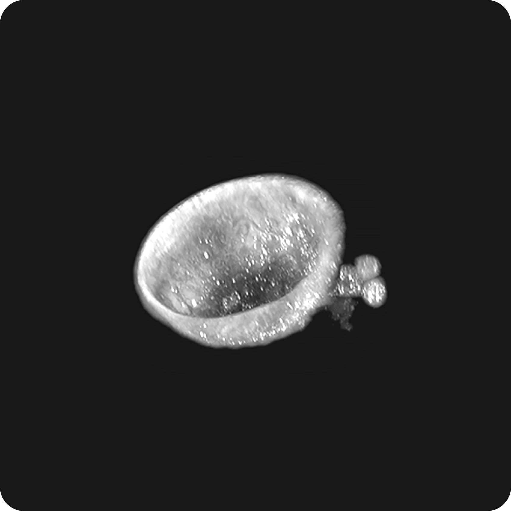
The HT-X1™ mini delivers similar high-resolution, label-free 3D imaging quality to the flagship in the HT-X1™ series — ensuring that every dataset is directly comparable across the HT-X1™ family. All analysis is powered by TomoAnalysis™, Tomocube’s AI-driven analysis platform. Researchers can perform advanced, automated workflows including nuclei detection, cell volume segmentation, and lipid droplet quantification. For mitochondria, the software provides detailed analysis of morphology and network connectivity, capturing structural changes that are critical for studying cellular dynamics. The software also supports various sample types such as organoids, blood cells, yeast, and bacteria, with options to integrate fluorescence imaging data. By combining consistent imaging with a unified analysis ecosystem, the HT-X1™ mini enables reproducibility, seamless collaboration, and confidence that every experiment meets the same high-quality standard.

-
Streamlined Workflow for Every User
The HT-X1™ mini keeps your imaging sessions fluid and uninterrupted. With a clear, step-by-step acquisition flow — load → focus → preview → capture — researchers can move efficiently from one sample to the next without repeated setup steps or system adjustments. Because holotomography is label-free, samples require no staining or washing, and can return directly to culture after imaging. This allows imaging to fit naturally into your routine — whether you're monitoring cells during multi-day experiments or capturing snapshots throughout the week. Once acquisition completes, 3D reconstruction begins immediately, so results are ready for review in the same workflow. The interface is intuitive and consistent, helping users stay focused on biology and experimental goals rather than system operation.
Applications


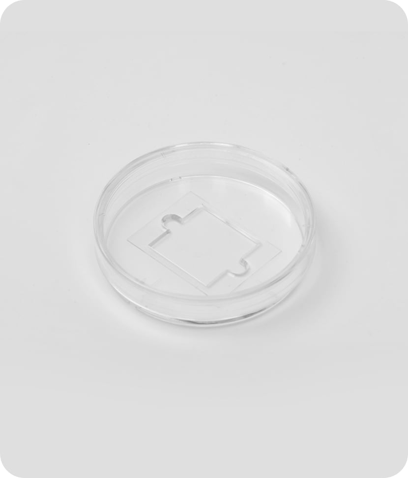
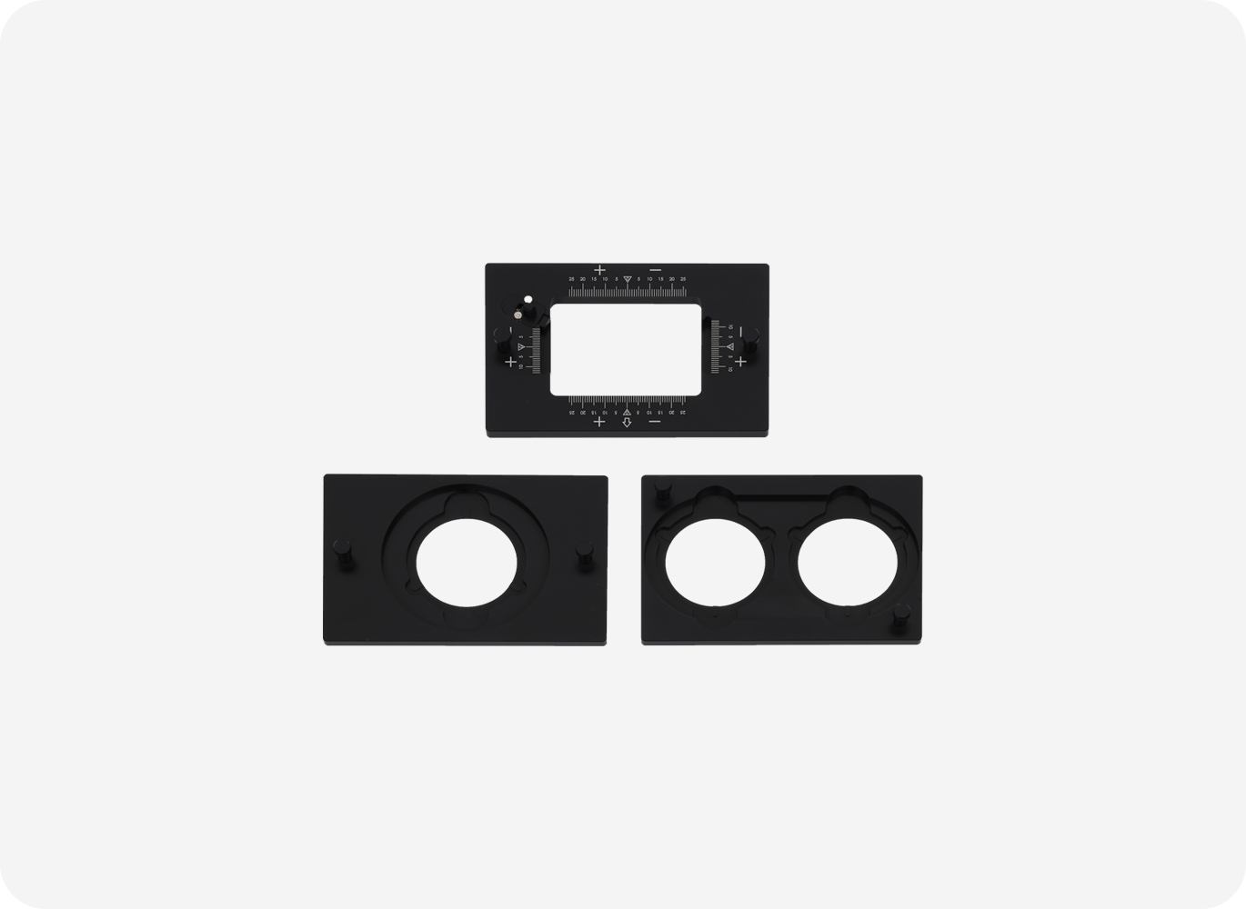
Configuration
-
HT-X1 mini main unit
- Objective Lens 40× NA 0.75 Air
- Image Sensor 2.8 MP CMOS
- Illumination LED 444 nm (520/660 nm optional)
- Motorized Stage
- Fluorescence Light engine (3-channel, Optional)
- Laser-based consistent focus (Optional)
- Environmental Controller (Optional)