
Regenerative medicine
Regenerative medicine, a dynamic field at the intersection of biology, engineering, and medicine, aims to restore or replace damaged tissues and organs by harnessing the body's natural healing processes. In this context, imaging and analysis play pivotal roles in understanding tissue regeneration, optimizing therapeutic interventions, and evaluating treatment outcomes.
The need for advanced imaging and analysis tools in regenerative medicine arises from the complex nature of tissue repair and regeneration processes. Traditional imaging techniques, often limited to two-dimensional views or requiring invasive labeling procedures, may not fully capture the intricacies of tissue dynamics. Meanwhile, Holotomography (HT), as an advanced label-free 3D live cell imaging technology, holds immense promise in regenerative medicine.
Unlike conventional imaging methods, HT provides non-invasive, high-resolution 3D imaging capabilities without the need for exogenous labels or stains. This enables researchers to visualize cellular spatial arrangement, extracellular matrix components, and cellular structures and dynamics in their natural state, preserving cell viability and physiological function.
HT's ability to quantify cellular features, such as volume, concentration, and dry mass, further enhances its utility in regenerative medicine. This quantitative analysis facilitates the characterization of engineered tissues, evaluation of therapeutic efficacy, and monitoring of tissue development over time.
Moreover, sample reusability in HT imaging is particularly advantageous in regenerative medicine research. By enabling repeated imaging of the same sample without altering its integrity, HT allows researchers to track longitudinal changes in tissue morphology and function. This longitudinal analysis is essential for understanding the temporal dynamics of tissue regeneration, optimizing treatment protocols, and assessing long-term outcomes.
In summary, the integration of HT into regenerative medicine research and clinical practice offers a powerful tool for advancing our understanding of tissue regeneration and enhancing therapeutic strategies. Its high-resolution 3D imaging, label-free, quantitative analysis capabilities, and ability to reuse samples make HT indispensable for visualizing and analyzing complex biological processes in regenerative medicine.
Features
Discover Regenerative medicine with HT
-
Cell therapy using CAR-T
Holotomography, an innovative imaging technology, has great potential in cell therapy, especially in the treatment of chimeric antigen receptor T cells (CAR-T). This technology provides valuable insights into cell behavior, quality, and function in both cell therapy research and clinical settings and goes a long way toward improving the clinical outcomes of cell-based therapy.
In cell therapy, Holotomography enables the detailed visualization and characterization of cell morphology, viability, and distribution. This provides important information necessary for quality control and optimization of cell manufacturing processes.
In addition, it can monitor cell behavior in real time, which helps to understand cell division, differentiation, and interaction with the surrounding environment, and to determine culture conditions for the optimal therapeutic effect. The quality and function of cells can be evaluated in a non-invasive and unlabeled manner, enabling fast and accurate quality control. This ensures consistency in cell therapy products.
In CAR-T treatment, CAR-T cell efficacy can be assessed, and treatment outcomes can be predicted by visualizing CAR-T cell morphology, active state, and interaction with target cells. In addition, by monitoring the expansion and proliferation of CAR-T cells in real time, the manufacturing process can be optimized to obtain the desired cell yield. The function of CAR-T cells can also be evaluated, which can be used to analyze target cell recognition, killing efficiency, cytokine secretion, etc., and improve CAR-T cell engineering strategies to improve treatment efficacy.
The utilization of Holotomography provides valuable insights into cell behavior, quality, and function in cell therapy research and clinical settings, and greatly helps to improve the clinical outcomes of cell-based therapy. -
Stem cell analysis & characterization
Observing living stem cells is critical for understanding their behavior and unlocking their therapeutic potential. By studying stem cells in their native state, researchers can assess their pluripotency and potential for differentiation, laying the groundwork for various applications in regenerative medicine and tissue engineering.
Additionally, monitoring living stem cells over time provides valuable insights into their dynamic interactions with the surrounding microenvironment and their response to external stimuli. This knowledge is instrumental in advancing our understanding of stem cell biology and harnessing its therapeutic potential for treating various diseases and injuries.
In a 2021 paper published in Molecules and Cells, researchers demonstrated that normal cells and stem cells can be distinguished based on their refractive index (RI) and volume in Holotomography (HT) images, eliminating the need for fluorescent markers (Kim et al., 2021).
This method effectively differentiated stem cells using RI and volume metrics, allowing quantification of nucleoli and vesicular structures unique to each cell type. Notably, stem cells exhibited significantly higher RI in vesicular structures compared to fibroblasts, as revealed by electron microscopy and HT analysis. These findings highlight HT’s potential to differentiate living stem cells from normal cells without fluorescence or specific biomarkers. -
Red blood cell research
Red blood cells (RBCs) research in regenerative medicine focuses on harnessing their unique properties for therapeutic purposes, particularly in tissue repair and regeneration. While RBCs are primarily recognized for their role in oxygen transport, they also possess characteristics that make them promising candidates for regenerative medicine applications.
Quantitative analysis of RBCs using holotomography offers a comprehensive approach to understanding their properties in the context of regenerative medicine. In a study published in Stem Cell Reports, researchers utilized HT to evaluate differentiated RBC characteristics derived from human induced pluripotent stem cells (hiPSC) (Sivalingam et al., 2021).
The 3D refractive index (RI) distributions of purified enucleated RBCs were obtained through HT, enabling the quantification of various morphological, biochemical, and mechanical properties. Morphological characteristics such as cellular volume, surface area, and sphericity were determined from reconstructed 3D RI tomograms.
Additionally, cellular hemoglobin concentration (CHC) and content (CH) were calculated based on the mean RI of the cell and the known refraction increment of hemoglobin. This information offered insights into the hemoglobin content of individual RBCs, essential for understanding their oxygen-carrying capacity and therapeutic potential. Dynamic mechanical properties of RBCs, specifically membrane fluctuations, were also assessed using HT. By generating 2D membrane fluctuation maps and analyzing temporal standard deviations of mean cell height profiles, researchers quantify the spatial average of membrane fluctuation over the projected cell area. This parameter provides valuable insights into the mechanical flexibility and integrity of RBC membranes, crucial for their function in circulation and potential involvement in regenerative processes.
Overall, HT-based analysis of RBCs offers a comprehensive understanding of their structural, biochemical, and mechanical properties, providing valuable insights for regenerative medicine applications. This approach enables the characterization of RBCs at a single-cell level, facilitating the exploration of their potential therapeutic roles in tissue repair and regeneration.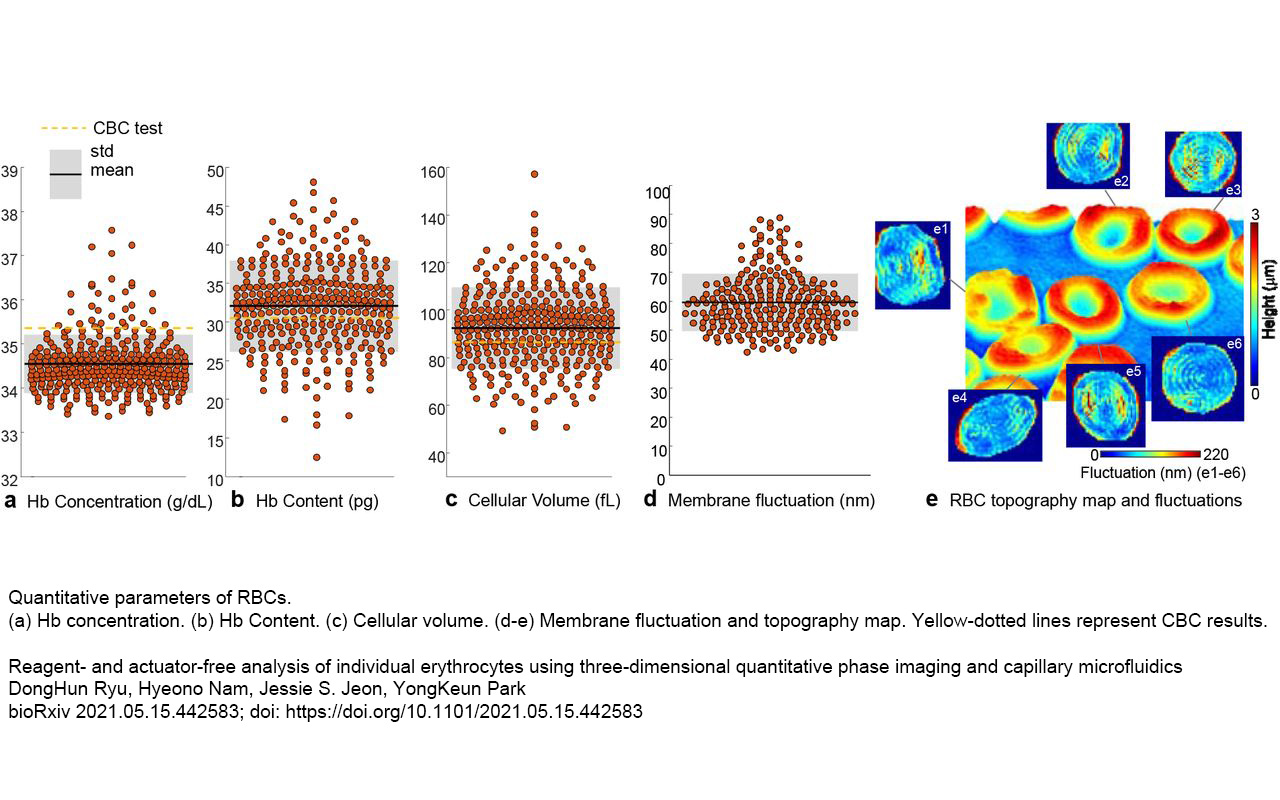
-
White blood cell research
White blood cells (WBCs) are integral to the immune system, defending against infections and diseases. The analysis of WBCs using HT technology holds promise for advancing regenerative medicine by enhancing our understanding of cell characteristics and interactions within the regeneration process. Such research endeavors are poised to contribute to the development of innovative treatments leveraging the regenerative potential of the immune system.
A recent study utilized Holotomography to assess the functionality of WBCs separated through different methods, revealing the advantages of using microfluidic as the separation technique (Lee et al., 2023). HT allows visualization of the 3D structure and internal composition of cells, aiding in understanding traits like WBCs’ morphology, motility, and phagocytic ability.
Timelapse imaging at high temporal resolution enabled the monitoring of phagocyte dynamics, while HT-fluorescence correlative imaging allowed for the examination and comparison of the phagocytic ability based on different separation strategies. The study suggested that HT was an optimal tool that minimized cellular deformation for image analysis in regenerative research.
Resources
Selected publications
-
Researchers demonstrated a suspension-culture-based differentiation process conducted under continuous agitation to scale up the entire procedure. HT was utilized to assess various cellular health and functional characteristics, such as cellular volume, surface area, hemoglobin content, hemoglobin concentration, and dynamic membrane fluctuations in purified populations of enucleated RBCs
-
Researchers differentiated normal cells and stem cells using their refractive index (RI) value and volume in Holotomography images, eliminating the need for fluorescent markers. This method quantified unique cellular structures, revealing higher RI in stem cell vesicular structures compared to fibroblasts. These findings highlight HT's potential to distinguish stem cells from normal cells without fluorescence or specific biomarkers.