3D biology
Biological research based on traditional 2D cell culture faces significant challenges regarding its inability to accurately mimic the complex spatial cellular organization and tissue dynamics found in vivo. Therefore, in recent years, the field of life sciences has undergone a swift transition towards 3D methods, fueled by advancements in 3D cellular models like spheroids and organoids or 3D cell culturing platforms like organ-on-a-chip. However, one of the primary technological challenges in 3D biology research lies in achieving high-resolution imaging while preserving the complex structure and integrity of the 3D culture models.
Within the realm of 3D biology imaging, the limitations of traditional imaging techniques such as confocal or fluorescence microscopies have become apparent as they either are unable to capture the complex details of individual cells in thick tissues, recapitulate the tissue-tissue interfaces, or often interfere with cellular functions and compromise data reliability by the use of exogenous agents for labeling or staining.
Meanwhile, Tomocube holotomography technology is the best suited for 3D biology research with a perfect balance among noninvasiveness, spatiotemporal resolution, signal-to-noise ratio, acquisition time, and simplicity.
First, Holotomography visualizes specimen details up to the subcellular level based on their refractive index. Without a labeling process, Holotomography preserves cell viability and allows for long-term monitoring of cellular dynamics within their 3D tissue environment. Second, LED-based non-interferometric optics have no speckle noise and have much faster scanning speeds. These features guarantee a spectacular high-contrast view of the dynamic complexity occurring inside 3D tissue models. Third, the method further allows quantitative measurement of 3D tissues and organoids, such as size, volume, mass, and growth rate, enabling a comprehensive, longitudinal tracking of the specimen characteristics.
Features
Discover 3D biology with HT
-
Organoids
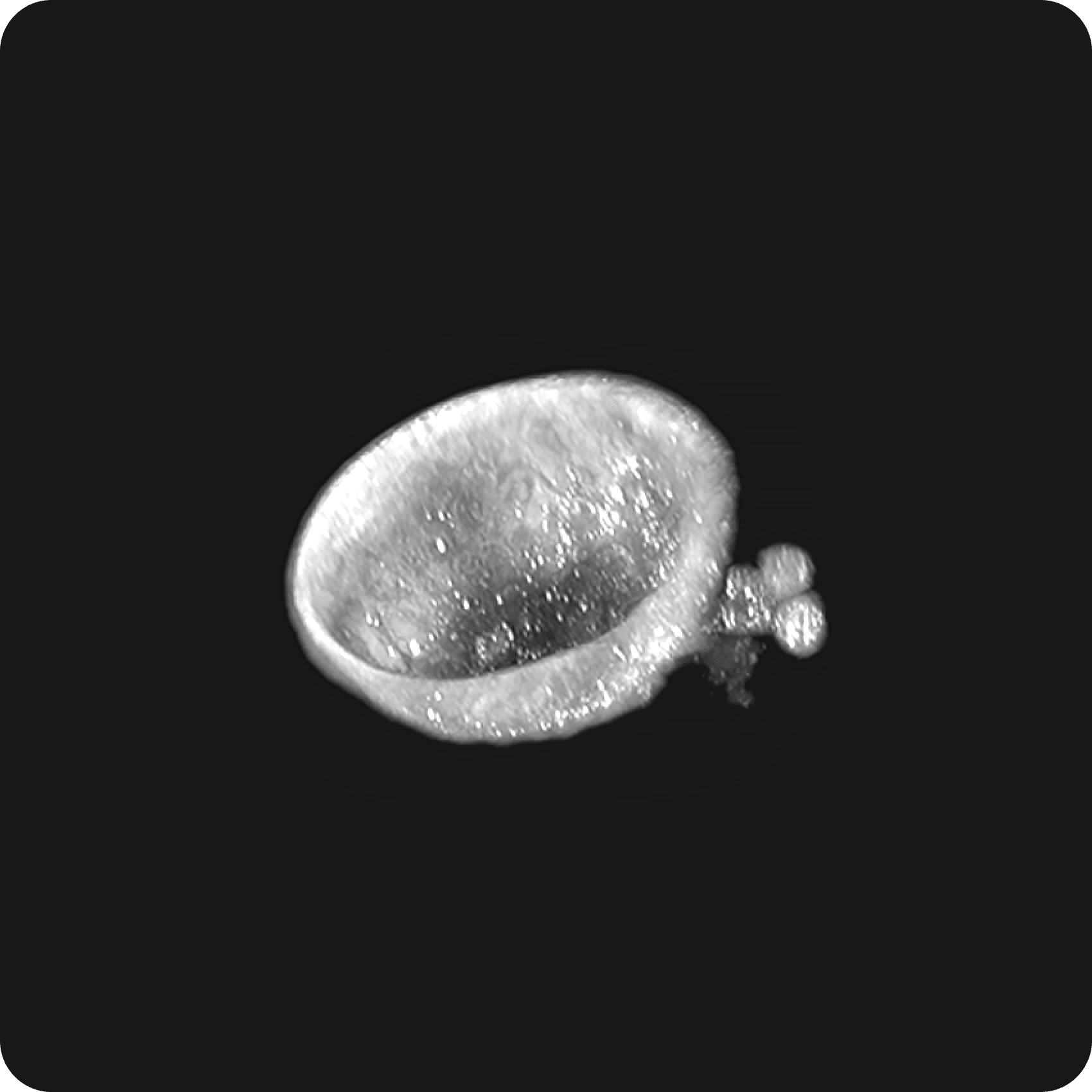
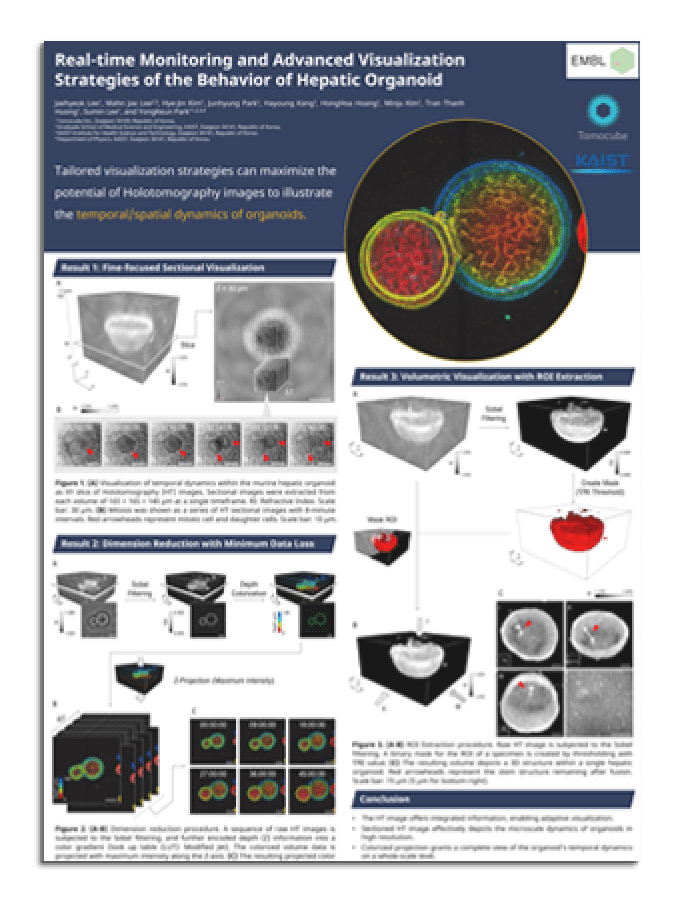
Organoids, the 3D cell aggregates replicating organ structures and functions, benefit significantly from HT's detailed and unlabeled imaging capabilities. It efficiently captures dynamic behavior of cells within living organoids, including mitotic events, epithelial cell dissection, cyst formation, and crypt budding. Real-time, high-resolution observations with available quantitative measurements facilitated by HT are crucial for scrutinizing the complex dynamics and morphology of the organoids.
In combination with the HT-ready 96 well plate, which supports a broad spectrum of cell types from 2D to 3D cultures, HT-X1 is an ideal tool to leverage the in vitro expandability of organoids to conduct high-throughput screenings for rapid assessment of drug response and its toxicity on tissues of individual patients.
In a recent study, HT was demonstrated to set a new standard for comprehensive, rigorous statistical assessments for the studies of organoids (Lee et al., 2023). With HT, intricate subcellular structures of individual cells in small intestinal organoids were observed in detail with a lateral resolution of 156 nm and axial resolution of 947 nm.
Long-term monitoring of different biological processes within the specimen, such as cell apoptosis, migration, mitosis, and other subcellular dynamics, could be easily achieved as the procedures required no time-consuming preparation or labeling.
In addition, observation of morphological changes was combined with quantitative parameters, including organoid volume, protein concentration, and mass, to effectively and comprehensively evaluate the response of the organoids to a chemotherapy drug treatment. The study suggested the Tomocube low-coherence HT imaging system as an indispensable tool for organoids in pharmacological screening applications. -
Advanced scaffold-based 3D cell cultures – Organ-on-a-chip
As the traditional 2D cell culture systems cannot mimic the natural tissue microenvironment due to the lack of cellular communication and interaction, it is essential to investigate the behavior of cells and tissues within a 3D context that can imitate the structure of the extracellular matrix (EMC) of the tissue.
Among several 3D cell culture techniques, microfluidic devices (µFDs) are in increasing demand for diverse biomedical research fields because of their ability to simplify and miniaturize integrated biological processes on microscale chips. µFDs are also proven to better mimic the dynamic mechanical properties and biochemical functionalities of the whole living organs than less advanced 3D culture systems, such as protein-based EMC or hydrogels.
However, conventional techniques face challenges in imaging miniaturized samples and labeling live specimens within µFDs due to limitations stemming from channel complexity and light scattering problems.
HT-X1 is a powerful tool that offers various benefits for observing cell dynamics in a 3D channel by providing a precise representation of cellular behavior in a 3D environment in high resolution and in real-time, enabling researchers to understand better how cells interact and behave in complex environments.
In addition, the HT-X1 can track cellular behaviors without the need for staining, which is useful when fluorescent staining is not easy for complicated 3D platforms. This capability allows researchers to observe cellular behavior without depending on staining techniques.
The excellent capturing ability of this low-coherence Holotomography system was demonstrated in an experiment aimed to observe live Hep3B cells residing on a 3D collagen type I and fiber structure within the DAX-1 chip.
High-resolution images of live Hep3B cells were achieved featuring detailed organelles structures including nucleus, nucleolus, lipid droplet, mitochondria, and collagen fiber at exceptional spatial resolution. Hep3B cells also displayed a distinct round-shape morphology that differs from their morphology in a 2D environment, highlighting the important influence of extracellular matrix (ECM) on cell behavior and morphology.
Resources
Selected publications
-
Research team proposed a new lung biopsy technique - cryobiopsy - resulting in a higher success rate for culturing lung cancer organoids (LCOs) while overcoming many critical limitations of conventional organoid models. Low-coherence HT was employed to evaluate the 3D structure and subcellular characteristics of LCOs without any pre-treatment. This proposed methodology promises to be a breakthrough strategy for the clinical application of LCOs in all stages of lung cancer.
-
HT sets a new standard for comprehensive, rigorous statistical assessments for the studies of organoids. With HT, researchers can observe intricate subcellular structures of individual cells in small intestinal organoids and easily perform long-term monitoring of different biological processes as the procedures required no time-consuming preparation or labeling. Observation of morphological changes was combined with quantitative parameters to effectively and comprehensively evaluate the response of the organoids to drug treatment.