Label-free quantitative imaging:
An innovative approach to exploring cells and tissues
-
- Holotomography(HT)
- HT illuminates a cell with very low power visible light at various illumination angles and measures the phase delay of the transmitted light. Although optically similar to CT, it differs in that it uses measured refractive index (RI) as imaging contrast, allowing visualization of living cells and tissues without labeling or staining.

Holotomography merges holography and tomography to solve the challenge of recovering light phase shifts to generate image contrast in Quantitative Phase Imaging, revealing intricate three-dimensional details within samples that would otherwise remain hidden with traditional techniques.
Just as a CT scan uses X-ray absorptivity as the imaging contrast to see inside a patient’s organs without invasive procedures, HT uses the refractive index (RI), an intrinsic optical parameter describing the speed of light passing through a specific material, to visualize living cells and tissues. Since variations in the biomolecular concentration directly impact the overall RI of the cellular biomaterials, the reconstruction of RI tomogram by Holotomography can retrieve important biophysical parameters such as cell volume, surface area, or protein concentration for further analysis.
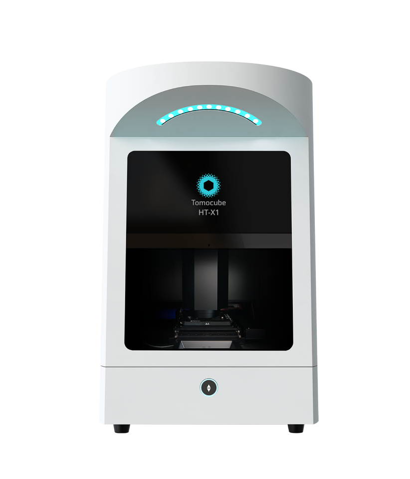
In general, all Holotomography systems consist of three essential components: (1) illumination optics to capture structural information of the specimen in light wave; (2) imaging optics to encode information in light onto a 2D intensity image; and (3) a reconstruction algorithm to decode and synthesize the raw data into a 3D tomogram.
Holotomography’s advantages lie in its capability to provide non-invasive, label-free 3D images of live cells, allowing real-time studies while the measurement of RI tomogram also allows quantitative analysis. Its data acquisition speed surpasses traditional methods, making it invaluable for quick results in medical diagnostics and drug research.
Holotomography’s applications span from cellular components like nuclei and mitochondria to studying organoids, tissues, and even whole organisms, enabling breakthroughs in cellular biology understanding. Its applications continue to expand as new advances are made in tomographic imaging.