Holotomography :
A Breakthrough in Industrial Metrology
-
- Holotomography(HT)
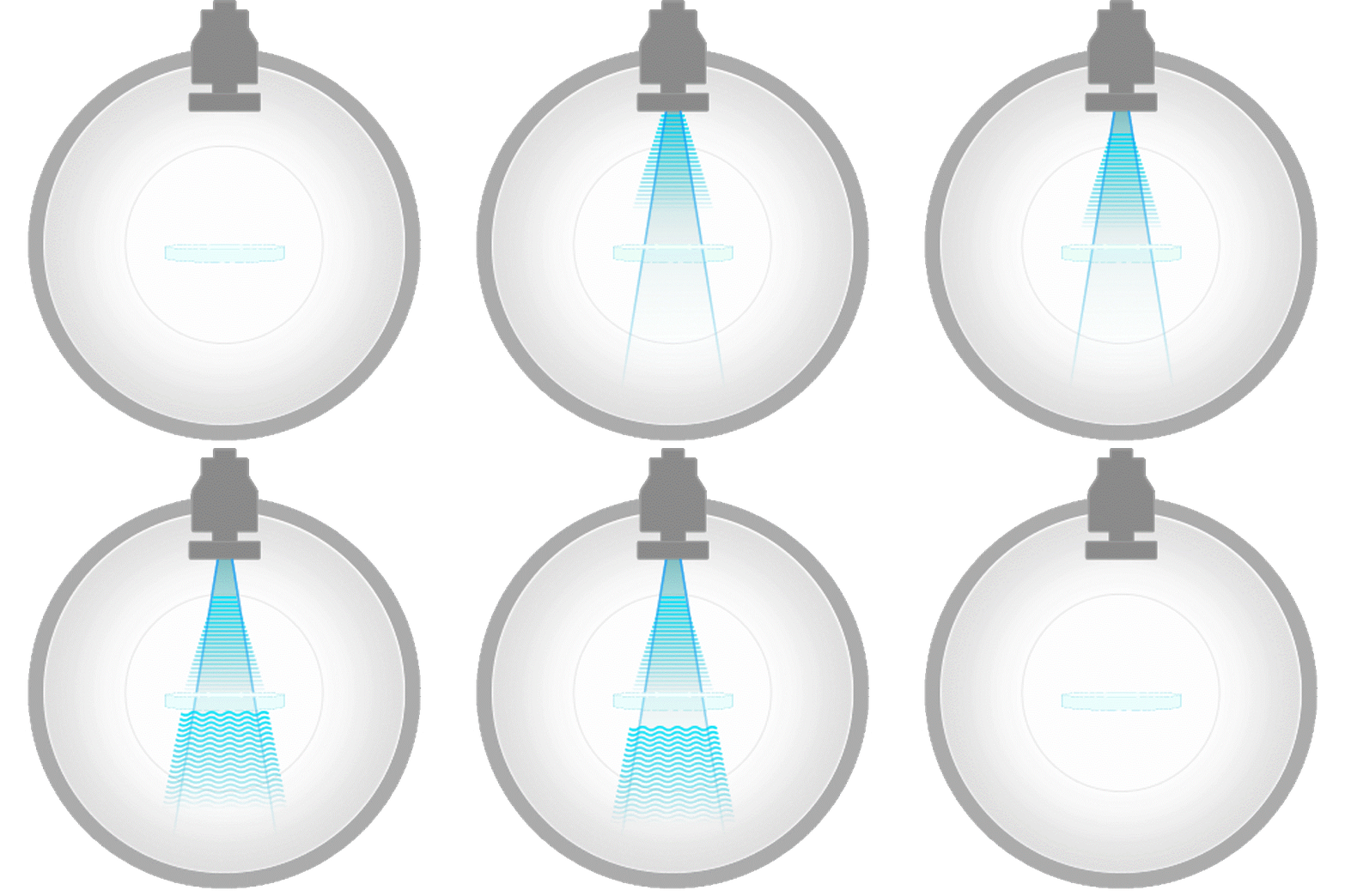
- Holotomography illuminates a sample from multiple angles and measures the phase delay of the reflected or transmitted light. While optically similar to CT, it differs by using measured refractive index (RI) variations as imaging contrast, enabling high-resolution visualization of transparent structures, multilayer thin films, and surface characteristics.
Next Generation 3D Imaging for Precision Inspection
Holotomography is a 3D optical imaging technique that combines holography and tomography to visualize internal structures of industrial materials with high resolution. By using quantitative phase imaging (QPI), it enables precise structural analysis beyond conventional metrology.
Like a CT scan reconstructs inner structures via X-ray absorption, holotomography maps refractive index (RI) distributions to create detailed 3D images. This provides valuable insights into transparent materials, multilayer thin films, step heights, and surface roughness.
Its advantages include nanoscale resolution (130–161 nm), non-destructive imaging, and quantitative measurements such as RI mapping, step height, and roughness. Real-time inspection is possible without damaging the sample, making it suitable for sensitive or complex materials.
Holotomography is highly compatible with various materials and is ideal for applications in semiconductors, glass substrates, optical elements, and electronic devices—expanding the scope of modern metrology and quality control.
Holotomography systems
How Holotomography Works
Holotomography is a "CT scan with light." Just as a hospital CT reconstructs the inside of the body from X-rays, holotomography reconstructs the inside of transparent materials — glass, polymers, optical films — from laser light, without cutting or coating the sample. Instead of X-ray absorption, it measures how much the light slows down at each point (the refractive index), revealing internal structures that ordinary microscopes simply cannot see.
Light passes through the sample
A low-power laser illuminates the sample. Wherever the material is denser, the light wave is delayed by a tiny, measurable amount — this delay is the "fingerprint" of the internal structure.
Holography records the delay
The delayed wave is compared against an undisturbed reference beam. Their interference pattern — a hologram — records the phase delay at every pixel with nanometer-scale precision.
Tomography rebuilds it in 3D
Holograms are captured from many illumination angles and computationally combined — just like CT — into a full 3D refractive-index map of the sample's interior, ready for measurement.
What the numbers mean: a lateral resolution of 130–161 nm is about 1/500th the width of a human hair, and a refractive-index sensitivity of 10⁻⁴ detects density changes far too subtle for conventional optical inspection.
How It Compares
Most fabs already own SEM, X-ray CT, or interferometry tools. Holotomography does not replace them — it fills the gap none of them covers: non-destructive, sub-micron 3D imaging of the inside of transparent materials.
| Capability | Holotomography | SEM cross-section | X-ray CT | White-light interferometry | Non-linear optical microscopy |
|---|---|---|---|---|---|
| Sees inside transparent materials | Yes — full 3D volume | Only at the cut plane | Weak contrast for glass / low-Z materials | No — surface only | Yes — full 3D volume |
| Destructive? | Non-destructive | Destructive (cutting / FIB) | Non-destructive | Non-destructive | Non-destructive |
| Lateral resolution | 130–161 nm | ~1–10 nm | ~0.5–1 µm class | ~0.3–0.5 µm class | ~0.5–1 µm class |
| Sample preparation | None | Sectioning, polishing, coating | Minimal | None | None |
| Typical measurement time for volume | Seconds – few minutes | Hours per cross-section | Tens of minutes – hours | Seconds – minutes | Over several minutes |
| Quantitative refractive-index map | Yes (Δn ~10⁻⁴) | No | No | No | No |
| Same sample re-usable after inspection | Yes — 100% intact | No | Yes | Yes | Yes |
* Representative values for typical tool classes, provided for orientation. Actual performance varies by model and sample. Contact us for an application-specific comparison.
Frequently Asked Questions
Q.What is Refractive Index (RI)?
Refractive index (RI) is a fundamental optical property that measures how much light is bent as it enters a material. It indicates the speed of light in a given medium compared to its speed in a vacuum. This property is determined by the material's optical density and molecular structure.
RI finds applications in various fields, including bioimaging, where its label-free and quantitative properties are particularly valuable. RI-based imaging techniques offer detailed insights into biological samples' structure and composition without the need for external labels or dyes. This makes them highly useful in biomedical research and diagnostics, providing high-resolution and sensitive imaging capabilities for studying cellular morphology, composition, and dynamics.
Q.What information can be acquired from holotomography?
Holotomography (HT), analyzed using software like TomoAnalysis, provides a wealth of information across multiple parameters. These include morphological, chemical, and mechanical aspects of the imaged samples.
Morphological Parameters: HT enables the measurement of various morphological parameters such as volume, surface area, projection area, and sphericity. These parameters offer insights into the shape and size characteristics of the imaged objects.
Chemical Parameters: Through holotomographic analysis, chemical information such as dry mass and concentration can be determined. For example, in the case of red blood cells, hemoglobin concentration can also be measured, providing valuable biochemical insights.
Mechanical Parameters: HT facilitates the measurement of mechanical properties such as cell stiffness. This parameter offers crucial information about the mechanical behavior and integrity of the imaged samples, which can be particularly relevant in studying cellular biomechanics and response to external stimuli.
Q.Does my sample need any preparation?
No. Holotomography is label-free and contact-free — no cutting, polishing, coating, or staining. Samples are measured as-is and remain fully intact afterwards, so the same part can continue through your process or be re-measured later.
Q.How long does a measurement take?
A single field-of-view acquisition takes seconds to minutes depending on volume depth and resolution settings. Larger areas are covered by automated stage stitching. For throughput estimates on your structure, request a sample measurement.
Q.What sample sizes and thicknesses can be measured?
Transmission systems (HT-T1™) image inside transparent materials such as glass up to 2 mm thickness. Stage travel and maximum panel size depend on configuration — contact us with your sample dimensions for a specific answer.
Q.Can it run in a production environment, or is it lab-only?
Both. The Module configurations are designed for integration into industrial inspection setups (with TomoPilot host interface), while Desktop configurations suit lab and pilot-line use with motorized stages and analysis software.
Q.How do I evaluate it with my own sample?
Send us a sample under NDA. We measure it free of charge, prepare a 3D analysis report, and review the dataset live with you. Use the Inquiry button or email precision@tomocube.com.
Q.What do I get as measurement output?
A full 3D refractive-index volume plus quantitative derived values — critical dimensions, depth and taper, step heights, surface and sidewall roughness, and defect locations — viewable and exportable in TomoAnalysis MI software.
See your own sample in 3D
The fastest way to evaluate holotomography is with your sample, not ours.
Send us a sample — we measure it and review the full 3D dataset with you, free of charge.
- 01Send your sample
under NDA - 02We measure it and prepare
a full 3D analysis report - 03Review the data live
with our application engineer